
Cesare Brizio
|
Fotomicrografia 2012/2013 |
|
Photomicrography 2012/2013 |
|
La Fotomicrografia, da non confondere con la microfotografia, è la fotografia di immagini ottenute utilizzando un microscopio. Solitamente, l'apparato fotografico prende il posto dell'oculare del microscopio. L'ingrandimento ottenuto dipende ovviamente dall'ottica del microscopio. Per fotografie digitali, entra in gioco anche l'ingrandimento digitale dipendente dal rapporto tra la dimensione del pixel sul sensore, e la dimensione del pixel sul monitor.
Per questi motivi, preferisco esprimere l'ingrandimento in termine di risolvenza (un sinonimo più elegante di "risoluzione"), indicando la risolvenza per ogni millimetro del soggetto. Photomicrography, not to be confused with microphotography, is photography of images obtained by using a microscope. Usually, the camera takes the place of the microscope's eyepiece. The available magnification of course depends on the microscope optics. For digital photos, digital magnification comes into play, that depends on the ratio between pixel size on the sensor, and pixel size on the monitor. For those reasons, I prefer to express magnification in terms of resolution, by indicating the resolution for each millimeter of the subject.
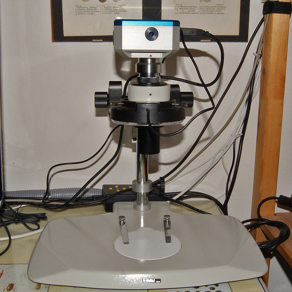
Ho acquistato l'equipaggiamento nel corso del 2012, e tra il 2012 e il 2013 ho realizzato le prime fotografie. Sono ancora ben lontano dall'avere risolto il problema chiave, quello della illuminazione. Ora dispongo di un illuminatore anulare a 144 led, a quattro zone, e sono in grado di fare solo fotografie in campo chiaro, con diversi problemi di illuminazione dello sfondo. Sebbene l'attrezzatura che possiedo non sia delle più costose, mi ha richiesto uno sforzo importante, e mi doterò con gradualità di strumenti professionali di illuminazione dello sfondo, valutando se ricorrere all'autocostruzione e se procurarmi un condensatore per microscopia in campo oscuro.
I purchased the equipment in 2012, and between 2012 and 2013 I took my first photographs. I'm still very far from having solved the key problem - lighting. Now I own a ring illuminator with 144 leds, four zones, and I'm able to do nothing but brightfield pictures, with several problems of background lighting. Although the equipment I own is not the most expensive, I took a major effort (around 3300 Euros), and I'll acquire very gradually a professional background lighting equipment, considering whether to resort to DIY and whether to acquire a condenser for darkfield microscopy.
Ho atteso l'esito dei concorsi Olympus Bioscapes e Nikon Small Word, ai quali ho partecipato senza alcuna speranza, prima di condividere con voi le mie immagini. Esse sono ottenute con la tecnica del "focus stacking": per mitigare l'impatto della scarsissima profondità di campo del microscopio, si effettuano numerose foto mentre l'altezza dell'obiettivo è progressivamente variata di frazioni di millimetro, in modo che ogni immagine abbia a fuoco una sottile "fetta" del soggetto. Una specie di tomografia assiale dell'esterno del soggetto. Software speciali, come Helicon Focus, usano la pila di immagini così ottenuta per selezionare solo le porzioni a fuoco di ogni scatto, utilizzate per comporre l'immagine risultante (una tecnica definita z-stacking). Per soggetti che eccedano l'inquadratura disponibile, il procedimento va ripetuto in più fasi, realizzando tante pile di immagini di differenti porzioni del soggetto, che viene traslato sotto il microscopio (in questo caso si parla di y-z stacking o di x-y-z stacking). Le diverse porzioni di immagine sono poi composte planarmente con software come Microsoft Image Composite Editor. I waited for the outcome of Olympus Bioscapes and Nikon Small Word competitions, in which I participated without any hope, before sharing my pictures with you. They are obtained by the technique of "focus stacking": to mitigate the impact of the microscope's extremely low depth of field, several shots are taken while the lens is progressively raised by fractions of a millimeter, so that each shot has a thin image "slice" in focus. Special software such as Helicon Focus, use the stack of images obtained to select only the best resolved portion of each picture, which are used to compose the resulting image (a technique called z -stacking). Sort of an axial tomography of the outside of the subject. For subjects that exceed the available field of view, the procedure is repeated in several stages, by creating several stacks of images from different portions of the subject, which is suitably translated under the microscope (in this case we speak of y-z stacking or x-y-z stacking). The different planar portions of the image are then composed by software such as Microsoft Image Composite Editor. Ho il sogno di riuscire a realizzare riprese di qualità di animali (Insetti e Ragni) vivi. Purtroppo, per ottenere un risultato adeguato bisogna trovare soggetti che restino perfettamente immobili per un tempo consistente (dai 5 ai 10 minuti), e che si prestino ad essere fotografati in studio (il mio equipaggiamento è molto scomodo da spostare e non si presta a un utilizzo in campagna). Non è semplice... qualche risultato preliminare mi incoraggia a proseguire.
I have a dream of being able to achieve high quality shots of alive animals (Insects and Spiders). Unfortunately, to obtain a proper result I should find subjects who remain perfectly still for a considerable time (from 5 to 10 minutes), and that lend themselves to be photographed in the studio (my equipment is very unpractical to move, and does not lend itself for field use). It is not easy ... some very preliminary results encourage me to continue.
Qui, propongo alcune foto dal mio primo anno di attività. Spero di potere migliorare col tempo, fino al punto di ottenere risultati di alta qualità. La fotomicrografia a basso ingrandimento rappresenta un'evoluzione della mia esperienza di macrofotografo, ed esplora una "terra di mezzo" fra la macrofotografia e la fotomicrografia spinta, documentando la bellezza di strutture di dimensione millimetrica. Tutte le mie foto sono rilasciate con licenza Creative Commons CC BY-NC-SA 3.0 Here, I propose some pictures from my first year of activity. I hope to improve over time, up to the point of obtaining high quality results. Low magnification photomicrography is an evolution of my experience as a macrophotographer, and explores a "middle-earth" between macrophotography and high magnification photomicrography, that can show the beauty of millimeter-size structures. All my pictures are released under Creative Commons CC BY-NC-SA 3.0 license. |
|
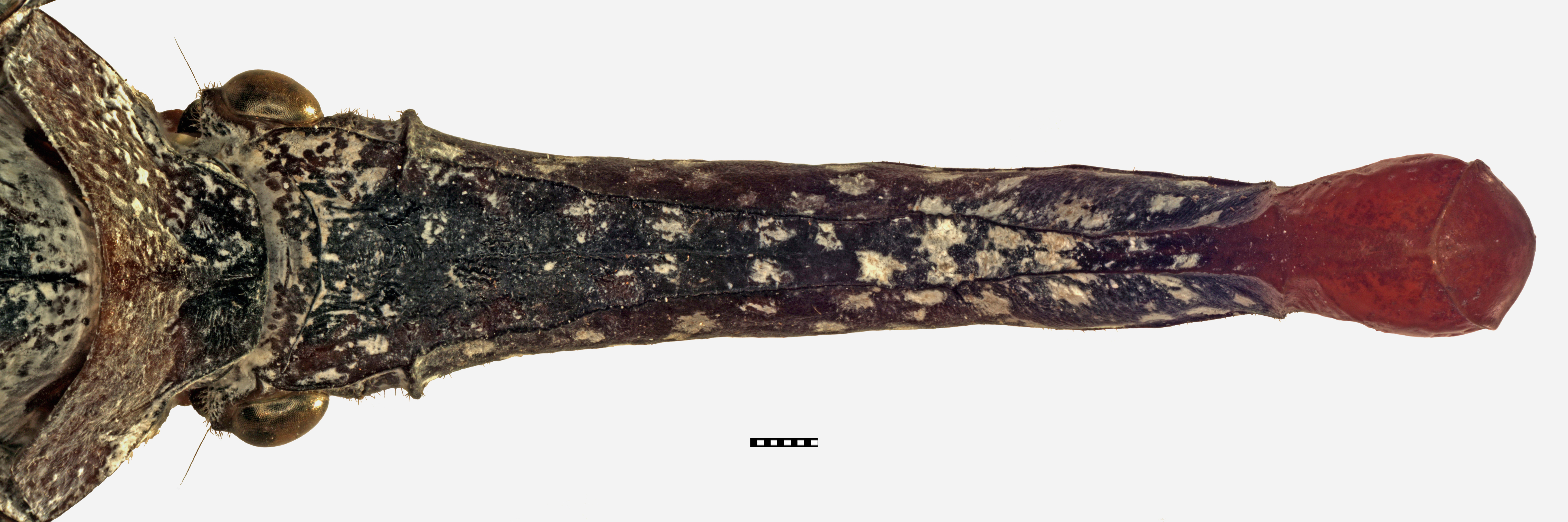
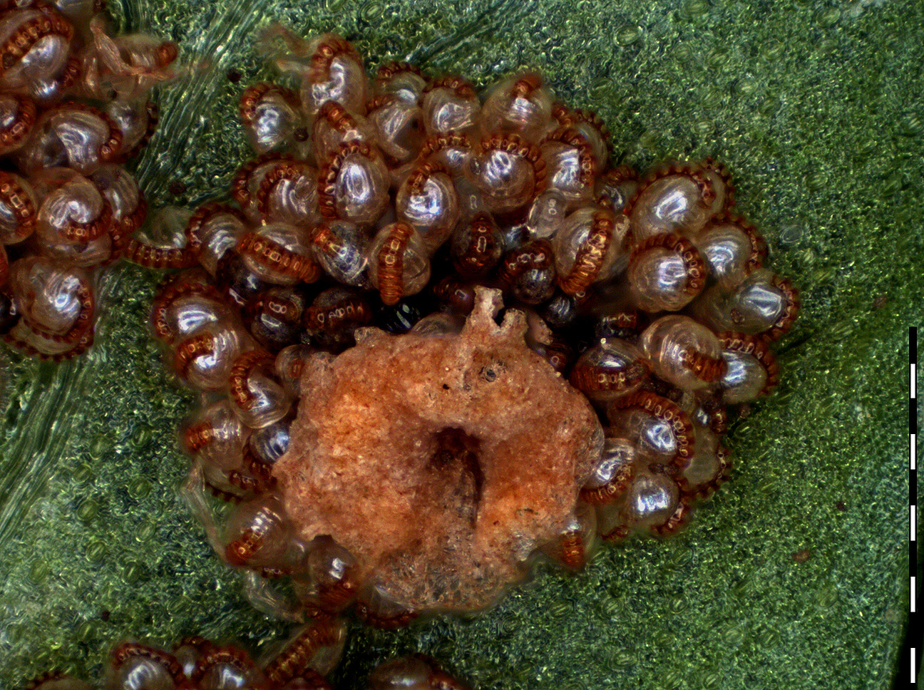
"Pinocchio"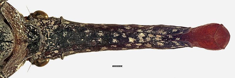
|
|
|
Risolvenza allo scatto: ~300 pixel/mm |
Resolution at capture: ~300 pixel/mm |
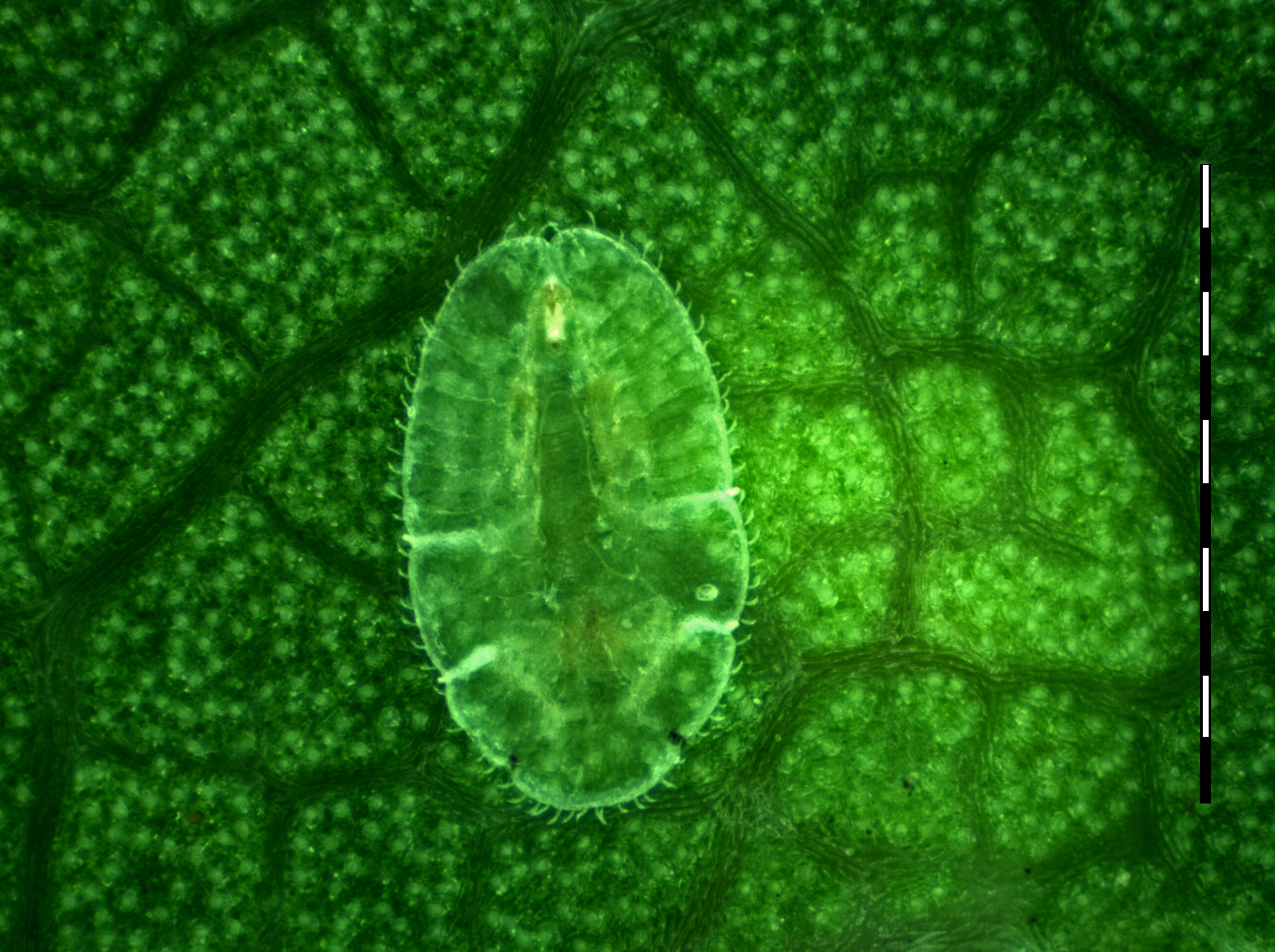
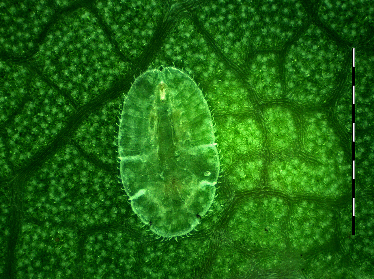
Natura morta in verde - Still life in green
|
|
|
Risolvenza allo scatto: ~1300 pixel/mm |
Resolution at capture: ~1300 pixel/mm |
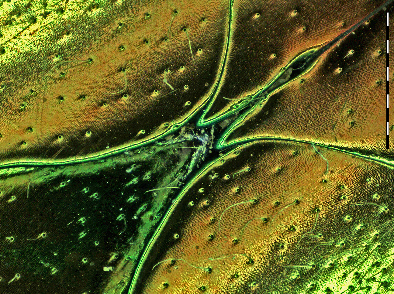
Convergenza - Convergence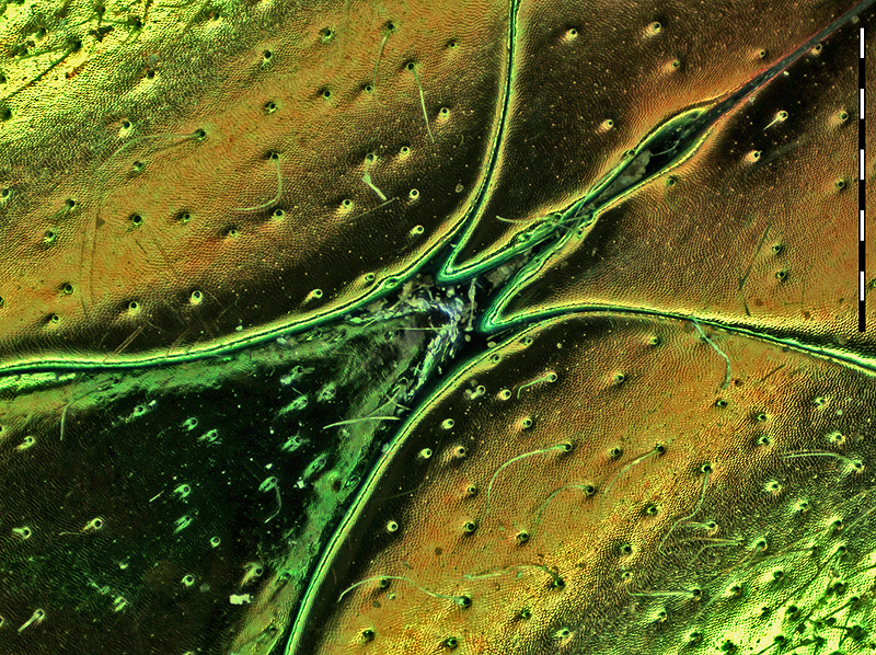
|
|
|
Risolvenza allo scatto: ~900 pixel/mm |
Resolution at capture: ~900 pixel/mm |
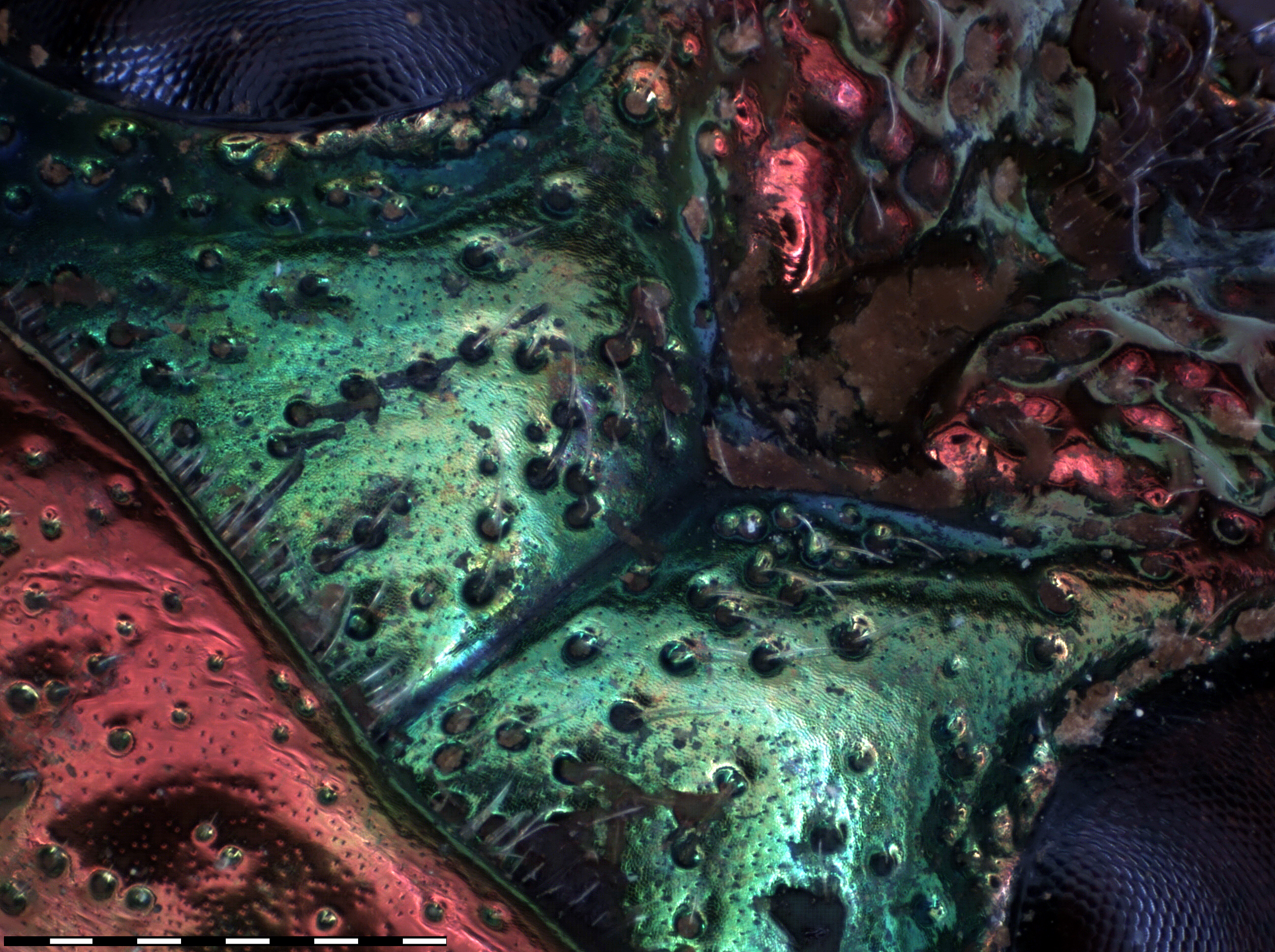
Una fronte aggrottata - A furrowed brow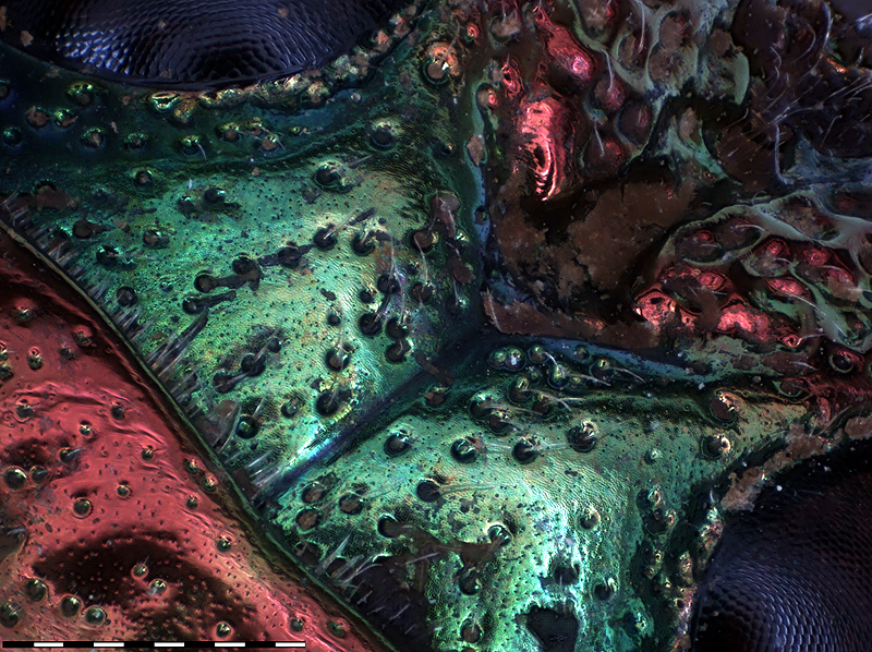
|
|
|
Risolvenza allo scatto: ~900 pixel/mm |
Resolution at capture: ~900 pixel/mm |
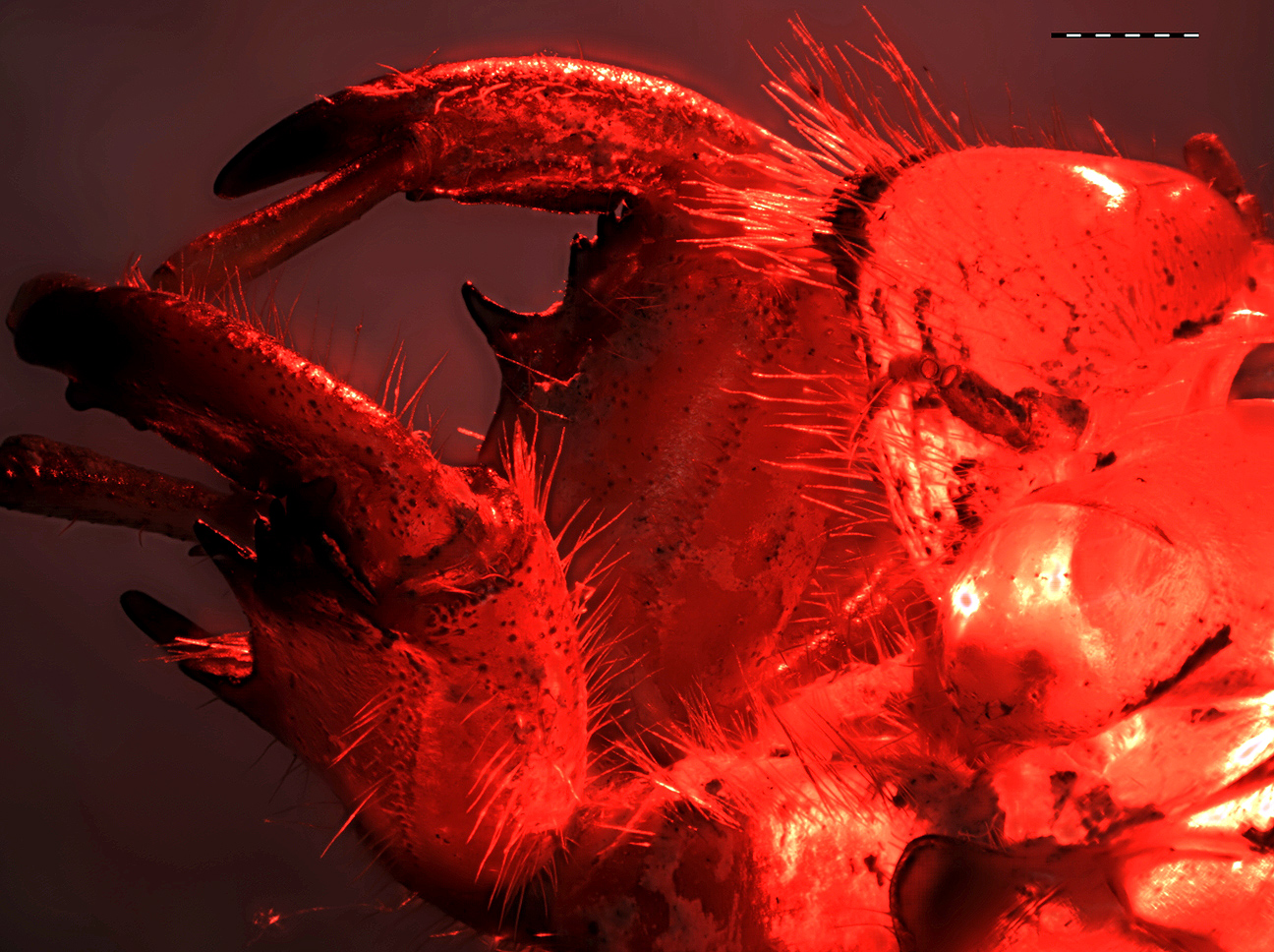
Fantasma Rosso - Red Phantom
|
|
|
Risolvenza allo scatto: ~300 pixel/mm |
Resolution at capture: ~300 pixel/mm |
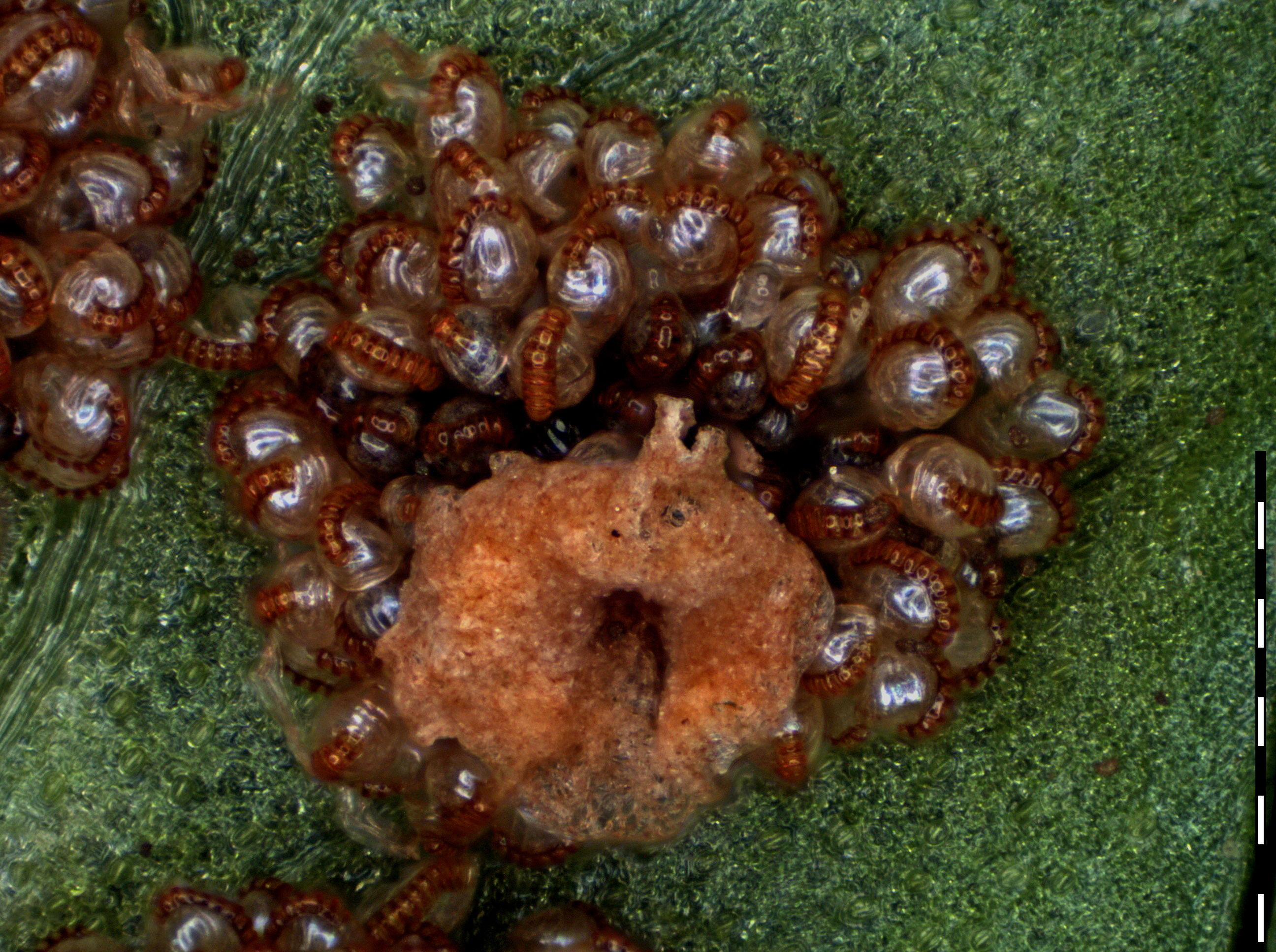
Gli occhi della tigre (un interessante fallimento) - The eyes of the tiger (an interesting failure)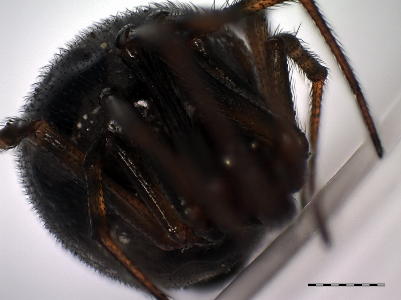
|
|
|
Risolvenza allo scatto: ~500 pixel/mm |
Resolution at capture: ~500 pixel/mm |
Felce natalizia - Christmas fern
|
|
|
Risolvenza allo scatto: ~1000 pixel/mm |
Resolution at capture: ~1000 pixel/mm |